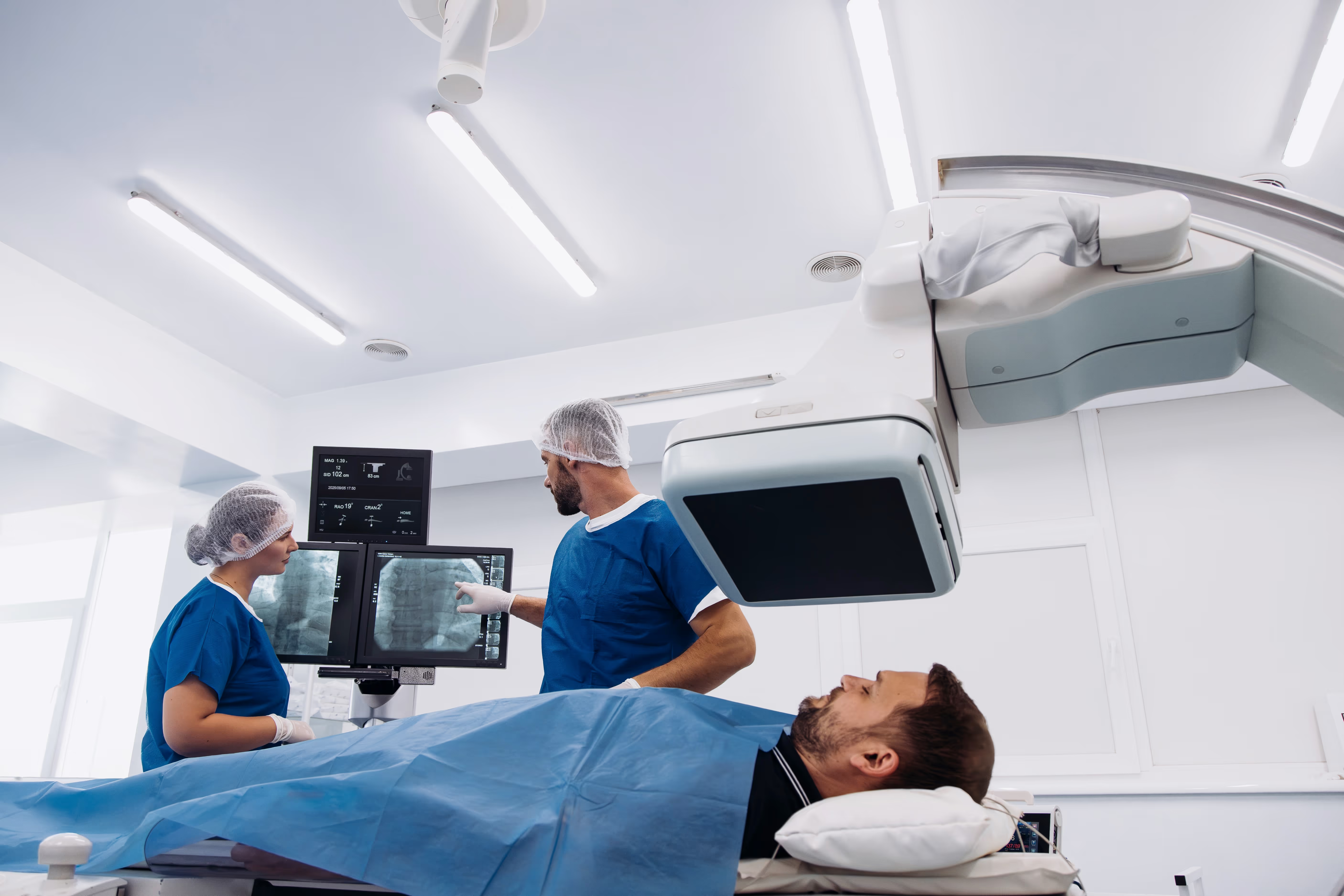
Minimally Invasive, Image‑Guided Care
Targeted Treatment. Lower Downtime.
Expert Interventional Radiologists
Precision Treatments with Less Downtime
Clear Support Before, During, and After
Advanced, Image‑Guided Procedures
What it is
Kyphoplasty is a minimally invasive procedure to treat painful vertebral compression fractures, often from osteoporosis. A small balloon is used to create space in the collapsed bone, then bone cement is injected to stabilize it.
Why it’s done
It can reduce back pain, improve mobility, and help restore vertebral height.
What to expect
Under imaging guidance, the radiologist places a small tube into the fractured vertebra, inflates a balloon, and injects cement. Sedation or anesthesia may be used. Most patients go home the same day.
Prep and safety
Tell us about blood thinners, infections, or allergies. Risks include bleeding, infection, cement leakage, or nerve irritation, which are uncommon. Specific preparation steps will be provided when your appointment is scheduled.
What it is
UFE is a minimally invasive treatment for symptomatic uterine fibroids. Tiny particles are delivered through a small catheter to block blood flow to fibroids, causing them to shrink.
Why it’s done
It helps relieve heavy bleeding, pelvic pain, pressure, and frequent urination without surgical removal of the uterus.
What to expect
A small puncture is made in the wrist or groin artery. Under X-ray guidance, the catheter is guided to the uterine arteries to deliver particles. Most patients go home the same day; cramping and fatigue are common for a few days.
Prep and safety
Not for those who are pregnant or have active infection. Discuss future fertility goals with your provider. Specific preparation steps will be provided when your appointment is scheduled.
What it is
PAE is a minimally invasive procedure for enlarged prostate (BPH). Tiny particles are used to reduce blood flow to the prostate, leading it to shrink.
Why it’s done
It can improve urinary symptoms like weak stream, urgency, and frequent nighttime urination, often with fewer sexual side effects than some surgeries.
What to expect
Through a small puncture in the wrist or groin, a catheter is advanced to the prostate arteries under X-ray guidance, and particles are delivered. Most patients go home the same day.
Prep and safety
Not suitable for everyone; imaging and lab tests may be needed beforehand. Risks include bruising, temporary urinary symptoms, or rare non-target embolization. Specific preparation steps will be provided when your appointment is scheduled.
What it is:
Hemorrhoid artery embolization is a minimally invasive procedure that treats internal hemorrhoids by reducing their blood supply. Using imaging guidance, a radiologist threads a tiny catheter into the arteries feeding the hemorrhoids and places microscopic particles to slow or block blood flow. With less blood flow, swollen hemorrhoids shrink and symptoms improve.
Why it’s done:
HAE is an option for people with bothersome bleeding, pressure, or prolapse from internal hemorrhoids who haven’t improved with diet changes, fiber, medications, or office procedures (like rubber band ligation). It can be an alternative to surgery for some patients, especially those seeking less pain and faster recovery.
What to expect:
After numbing the skin, a small catheter is placed through a wrist or groin artery. Under X‑ray guidance, the catheter is guided to the rectal arteries, and tiny particles are delivered. Most patients go home the same day. Soreness or pelvic pressure can occur for a few days and is usually managed with over‑the‑counter pain relief. A follow‑up visit checks your progress and next steps, if needed.
Prep and safety:
Tell us about blood thinners, allergies, pregnancy status, and any rectal bleeding history or prior surgeries. Imaging or labs may be needed beforehand. Risks are uncommon but can include bruising, temporary rectal discomfort, or non‑target embolization. Specific preparation steps will be provided when your appointment is scheduled.
What it is
PAD occurs when arteries in the legs narrow due to plaque buildup. We provide imaging to diagnose PAD and minimally invasive treatments to improve blood flow.
Why it’s done
To evaluate leg pain with walking, non-healing wounds, or cold, pale feet, and to prevent complications.
What to expect
Tests may include ultrasound, CT angiography, MR angiography, or catheter-based angiography. Treatments can include balloon angioplasty, stenting, or atherectomy, done through a small puncture with X-ray guidance.
Prep and safety
Tell us about kidney issues, diabetes, and blood thinners. Contrast dye may be used. Specific preparation steps will be provided when your appointment is scheduled.
What it is
DVT is a blood clot in a deep vein, usually in the leg. We use imaging to diagnose DVT and, in select cases, provide minimally invasive treatments.
Why it’s done
Prompt diagnosis helps prevent a clot from traveling to the lungs (pulmonary embolism) and reduces long-term vein damage.
What to expect
Ultrasound is the main test. Some patients may need CT or MR venography. Treatment options can include blood thinners and, in certain cases, catheter-directed thrombolysis or thrombectomy to remove or dissolve the clot.
Prep and safety
Share your medication list and bleeding history. Urgent evaluation is important for new leg swelling or pain. Specific preparation steps will be provided when your appointment is scheduled.
What it is
Varicocele embolization treats enlarged veins in the scrotum (varicoceles) by blocking abnormal blood flow with tiny coils or medication.
Why it’s done
It can reduce pain, shrink the varicocele, and may improve fertility in some patients.
What to expect
Through a small vein puncture, a catheter is guided to the affected veins under X-ray. Coils or sclerosant are placed to seal them. Most patients go home the same day.
Prep and safety
Mild soreness or bruising can occur. Rare risks include vein irritation or migration of material. Specific preparation steps will be provided when your appointment is scheduled.
What it is
PCS is chronic pelvic pain caused by enlarged pelvic veins. Embolization can seal these veins to reduce pain.
Why it’s done
To treat daily pelvic aching, pain that worsens standing or after intercourse, and related symptoms when other causes have been ruled out.
What to expect
A catheter is placed through a small vein puncture and guided to the ovarian or pelvic veins. Coils or medication close off the problem veins. Most patients return to routine activities in a few days.
Prep and safety
Tell us if you are pregnant or trying to conceive. Risks are low but include bruising and temporary discomfort. Specific preparation steps will be provided when your appointment is scheduled.
What it is
Minimally invasive treatments use imaging guidance to target tumors. Options may include ablation (freezing or heating), embolization, or radioembolization.
Why it’s done
These therapies can control or shrink tumors, relieve symptoms, and work alongside surgery, chemotherapy, or radiation.
What to expect
Depending on the plan, a needle or catheter is used to reach the tumor with imaging guidance. Treatments are tailored to tumor type and location, often with short hospital stays.
Prep and safety
Pre-procedure labs and imaging are required. Tell us about medications, especially blood thinners, and any liver or kidney issues. Specific preparation steps will be provided when your appointment is scheduled.
What it is
GAE is a minimally invasive procedure to treat chronic knee pain from osteoarthritis by reducing abnormal blood flow and inflammation in the knee lining.
Why it’s done
It can lessen pain and improve function for patients who are not ready for or cannot undergo knee replacement, or who have persistent pain after other treatments.
What to expect
Through a small wrist or groin puncture, a catheter is guided to the knee arteries and tiny particles are delivered. Most patients go home the same day.
Prep and safety
Temporary skin warmth or mild discomfort around the knee can occur. Not for everyone; evaluation is required. Specific preparation steps will be provided when your appointment is scheduled.
What it is
IVC filter retrieval is a minimally invasive procedure to remove a small metal device (inferior vena cava filter) from the large vein in your abdomen. Filters are placed to catch blood clots traveling from the legs to the lungs. When the risk of clots has passed, removing a retrievable filter helps lower long‑term risks like filter fracture, movement, or vein blockage.
Why it’s done
Filters are meant to be temporary for many patients. Retrieval is recommended when you can safely use blood thinners again, your clot risk has decreased, or your care team decides the filter is no longer needed. Taking it out reduces the chance of future complications and makes future imaging and treatments easier.
What to expect
You will lie on the procedure table while the skin is cleaned and numbed. A small tube (catheter) is placed through a neck or groin vein. Using X‑ray guidance, the radiologist gently grasps the filter with a snare or special device and removes it through the catheter. Most removals take 30–60 minutes, and many patients go home the same day. If the filter has been in place a long time or is tilted or embedded, advanced tools may be used, which can take longer.
Prep and safety
Tell us about blood thinners, allergies (especially to contrast dye), kidney problems, and any prior filter procedures. Imaging and lab tests may be needed before the retrieval. Risks are uncommon but can include bleeding, infection, vein injury, or rare filter breakage. Specific preparation steps will be provided when your appointment is scheduled.
What it is
Arterial aneurysm repair treats a weakened, bulging section of an artery—most commonly in the aorta or in leg arteries—to prevent rupture or reduce symptoms. Using imaging guidance, a radiologist places a fabric-covered metal tube (stent graft) inside the artery through small punctures, creating a new inner lining so blood flows safely and no longer presses on the aneurysm wall.
Why it’s done
Repair lowers the risk of life‑threatening rupture and can relieve pain or pressure from large or fast‑growing aneurysms. It is often recommended when an aneurysm reaches a certain size, grows quickly, or causes symptoms. Endovascular repair can be an option for many patients who may not be candidates for open surgery.
What to expect
You’ll lie on a procedure table while the skin in the groin (and sometimes wrist) is cleaned and numbed. Under X‑ray guidance, small tubes are guided into the artery and the stent graft is positioned across the aneurysm, then expanded to fit snugly. Most patients spend one night in the hospital and return to light activity within days.
Prep and safety
Tell us about blood thinners, allergies (especially to contrast dye), kidney problems, and prior vascular surgeries. Pre‑procedure CT or ultrasound and lab tests help plan the repair. Risks are uncommon but can include bleeding, infection, vessel injury, endoleak (blood still reaching the aneurysm sac), kidney strain from contrast, or need for future adjustments. Specific preparation steps will be provided when your appointment is scheduled.
What it is
Tumor embolization is a minimally invasive treatment that targets tumors by blocking their blood supply and delivering therapy directly where it’s needed. Two common types are:
TACE (Transarterial Chemoembolization): mixes chemotherapy with tiny particles to deliver medicine into the tumor’s artery and slow its blood flow.
Y-90 Radioembolization: delivers microscopic beads loaded with yttrium-90 (a low‑penetration radiation source) into the tumor’s artery to radiate it from the inside.
These treatments are most often used for liver tumors, including hepatocellular carcinoma and certain metastases, but may be used in other locations based on your care plan.
Why it’s done
By treating the tumor from the inside, embolization can shrink or control tumors, relieve symptoms, and help bridge or downstage patients to surgery or transplant. It can also be used when surgery or systemic therapy alone is not the best option.
What to expect
A small puncture is made in the wrist or groin artery. Using X‑ray guidance, the radiologist guides a catheter into the arteries feeding the tumor. The treatment—TACE medicine or Y‑90 beads—is then delivered. Most patients go home the same day or after an overnight stay. With TACE, you may have temporary fatigue, fever, nausea, or right‑upper‑belly soreness (called post‑embolization syndrome) for a few days. Y‑90 side effects are often milder but can include tiredness and brief discomfort. Follow‑up imaging checks response and plans next steps.
Prep and safety
Pre‑procedure planning often includes blood tests and detailed imaging (CT/MRI). For Y‑90, a separate mapping angiogram is typically done first to plan dose and protect nearby organs. Tell us about blood thinners, allergies (especially to contrast dye), kidney or liver problems, and if there’s any chance of pregnancy. Risks are uncommon but can include bleeding, infection, non‑target embolization, or liver irritation. Specific preparation steps will be provided when your appointment is scheduled.
What it is
Tumor ablation is a minimally invasive treatment that destroys tumors by applying extreme heat or cold through a thin needle placed directly into the tumor using imaging guidance. Common methods include:
Radiofrequency or microwave ablation (uses heat to destroy tumor cells)
Cryoablation (uses freezing to destroy tumor cells)
These techniques are often used for small tumors in the liver, kidney, lung, or bone, and can be repeated if needed.
Why it’s done
Ablation can control or eliminate tumors while preserving healthy tissue. It may be used when surgery isn’t the best option, to treat limited new spots, to relieve pain (especially in bone), or alongside other therapies such as chemotherapy, targeted therapy, or radiation.
What to expect
You will lie on a procedure table while the skin is cleaned and numbed. Most ablations use sedation or anesthesia for comfort. Using CT, ultrasound, or X‑ray guidance, the radiologist places one or more thin probes into the tumor and applies heat or cold for several minutes. You may stay for a few hours of observation or overnight, depending on the location and size treated. Follow‑up imaging is done to confirm the result and guide any additional treatment.
Prep and safety
Tell us about blood thinners, bleeding disorders, lung disease, kidney or liver problems, and any implanted devices. Risks vary by organ but can include bleeding, infection, damage to nearby structures, or temporary pain/swelling. Lung ablation has a small risk of air leak (pneumothorax), and liver ablation can cause temporary liver irritation. Specific preparation steps will be provided when your appointment is scheduled.

Our Providers
Find a Location