Breast Imaging with Clarity and Care
Early Answers. Calm Guidance.
Expert Breast Imaging Specialists
Clear, Easy‑to‑Understand Results
Modern Imaging for Early Detection
Your Breast Health, Clearly Explained
What it is:
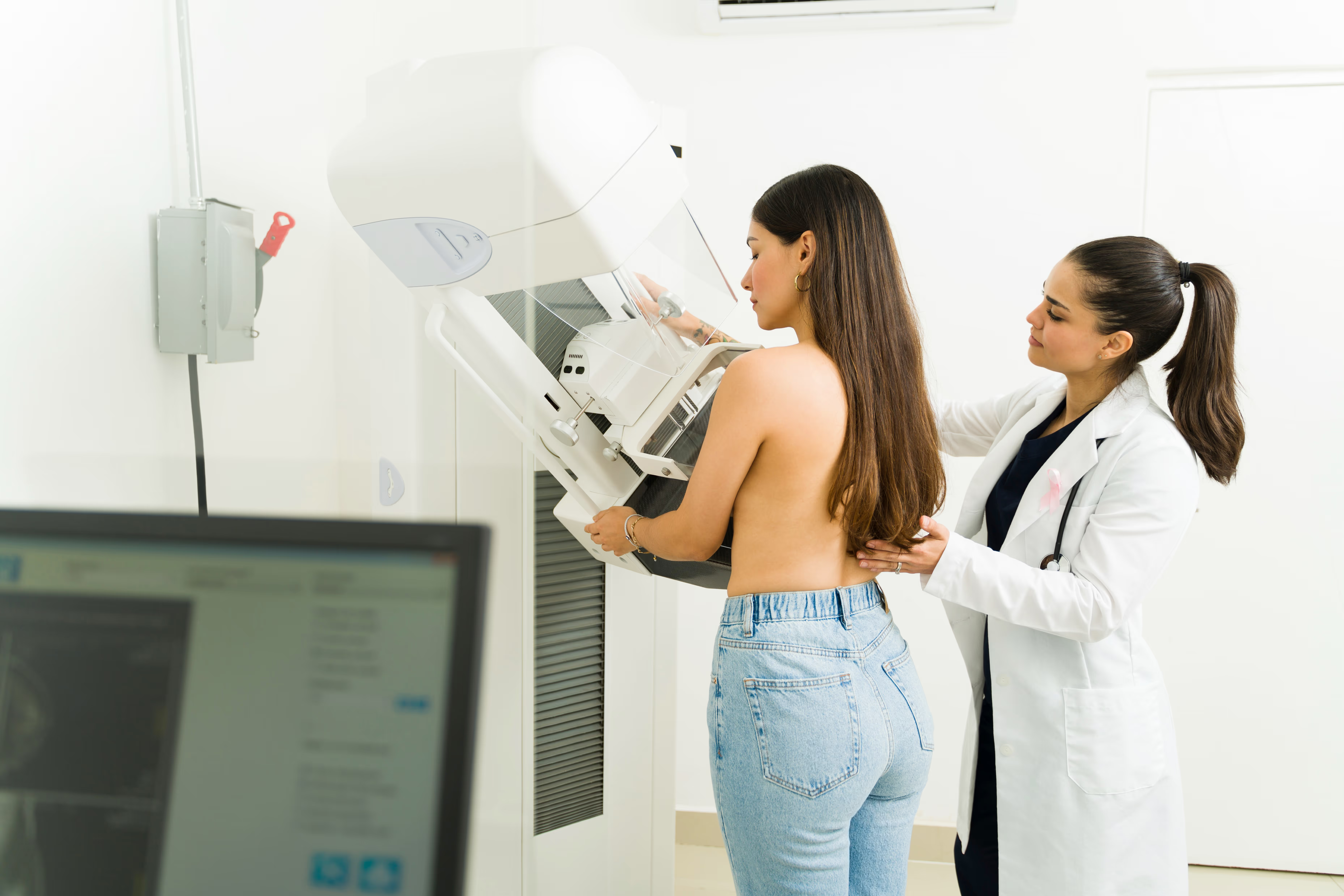
A mammogram is a low-dose X-ray of the breast. It helps find breast changes, including cancers, that may be too small to feel.
Why it’s done:
Mammograms are used to screen for breast cancer and to check new symptoms like a lump, pain, or nipple changes. They help your care team decide next steps.
What to expect:
Your breast is placed on a flat plate and gently compressed for a few seconds while images are taken. You may feel pressure, but it should be brief. A technologist positions you for several views. The exam usually takes 15–30 minutes.
Prep and safety:
Avoid deodorant, powders, or lotions under your arms or on your chest the day of the exam, as they can show up on images. Tell us if you are pregnant or breastfeeding, or if you have breast implants. Specific preparation steps will be provided when your appointment is scheduled.
What it is
A screening mammogram is a routine X-ray test for people without breast symptoms. It looks for early signs of cancer before they cause problems.
Why it’s done
Regular screening lowers the chance of dying from breast cancer by finding it early, when it’s most treatable. It is typically done once a year based on age and risk.
What to expect
Both breasts are imaged with gentle compression in several views. The visit usually takes 15–20 minutes. If anything looks unclear, you may be called back for extra images or an ultrasound.
Prep and safety
Do not use deodorant or powders on the day of imaging. Bring prior mammograms if done elsewhere. Let us know about implants or prior breast surgery. Specific preparation steps will be provided when your appointment is scheduled.
What it is
A diagnostic mammogram is a targeted breast X-ray used to evaluate symptoms (like a lump or discharge) or to study an area seen on a screening exam.
Why it’s done
It provides extra, detailed views to help clarify a finding and guide next steps, such as ultrasound, MRI, or biopsy if needed.
What to expect
The technologist will take additional angles and magnified views of the area of concern. An ultrasound may be performed during the same visit. A radiologist reviews the images and discusses recommended next steps.
Prep and safety
Avoid deodorant or powders. Bring prior imaging for comparison if available. Tell us if you are pregnant, breastfeeding, or have implants. Specific preparation steps will be provided when your appointment is scheduled.
What it is
ABUS uses sound waves and a wide, automated scanner to create 3D images of the entire breast, especially helpful in people with dense breast tissue.
Why it’s done
Dense breasts can make cancers harder to see on mammograms. ABUS adds another look to improve detection alongside screening mammography.
What to expect
You lie on your back while a layer of gel is applied. A curved probe gently scans each breast in minutes, without radiation or compression. A radiologist interprets the images after the exam.
Prep and safety
Wear a two-piece outfit for comfort and avoid heavy lotions on the chest. Safe in pregnancy since there is no radiation. Specific preparation steps will be provided when your appointment is scheduled.
What it is
Breast ultrasound uses sound waves to create pictures of breast tissue. It helps tell if a lump looks solid or fluid-filled (a cyst).
Why it’s done
It evaluates symptoms or findings from a mammogram and can guide procedures like needle biopsies.
What to expect
You lie on the exam table while a technologist moves a small handheld probe over the breast with gel. There is no radiation and it is usually painless. The exam takes about 15–30 minutes.
Prep and safety
No special prep. Safe during pregnancy and breastfeeding. Specific preparation steps will be provided when your appointment is scheduled.
What it is
Breast MRI uses a strong magnet and radio waves to make detailed images of the breast. Often contrast dye is used to highlight blood flow.
Why it’s done
It is used for high-risk screening, to define the extent of known cancer, to check implants, or to evaluate unclear findings from other tests.
What to expect
You lie face down on a padded table with openings for the breasts. The table slides into the MRI scanner. You’ll hear tapping sounds and should stay still. If contrast is used, it is given through an IV. The exam usually takes 30–45 minutes.
Prep and safety
Tell us about any kidney problems, metal implants, or a history of contrast reactions. Let us know if you are pregnant or may be claustrophobic. Specific preparation steps will be provided when your appointment is scheduled.
What it is
A needle biopsy uses imaging—such as ultrasound, mammography (stereotactic), or MRI—to guide a small needle into a targeted area to collect tissue samples.
Why it’s done
It helps determine if a finding is benign or cancerous without surgery, guiding treatment decisions.
What to expect
The skin is cleaned and numbed. Using real-time imaging, the radiologist places the needle to sample the area. You may feel pressure, but it’s usually well tolerated. A small marker may be left to mark the spot for future imaging. Most patients resume normal activity the same day.
Prep and safety
Tell us about blood thinners, bleeding disorders, or allergies to local anesthetics or contrast. Expect minor bruising or soreness. Specific preparation steps will be provided when your appointment is scheduled.

Our Providers
Find a Location